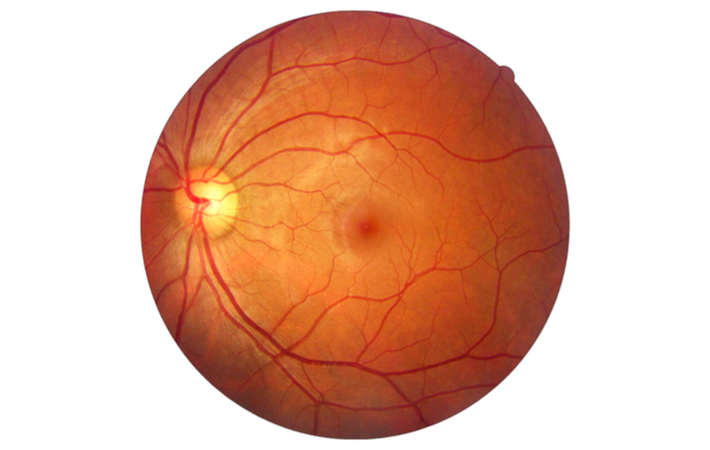
The Vitereo retina is a thin layer of tissue that lines the back of the eye on the inside. It is located near the optic nerve.
The purpose of the retina is to receive light that the lens has focused, convert the light into neural signals, and send these signals on to the brain for visual recognition.
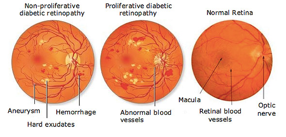
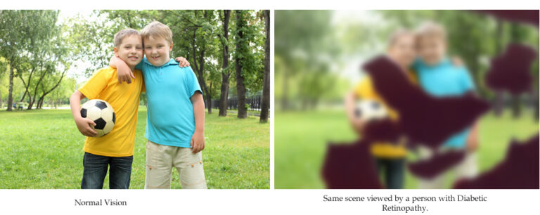
1. DIABETIC RETINOPATHY
Diabetic Retinopathy is the “disease of the retina” caused by microangiopathy due to long term effects of diabetes leading to progressive damage of the retina & blindness. The most common cause of severe bilateral visual loss in the working age group.
TREATMENTS
Laser Treatment: The aim of this treatment is to protect central vision. It does not restore lost vision, but it can prevent further deterioration, which is why early diabetic retinopathy diagnosis through periodic eye examination is imperative.
Intravitreal Injections (Anti VEGF agents): Anti VEGF agents (Lucentis, Avastin, Eyelea etc.)emerging as the new modality of treatment for various stages of diabetic retinopathy. These agents are injected into the eye (intravitreal injection). They are commonly used in diabetic maculopathy and proliferative diabetic retinopathy.
Surgery (VITRECTOMY): A microsurgical procedure where the blood filled vitreous gel is replaced with a clear gas/solution/oil to restore vision.
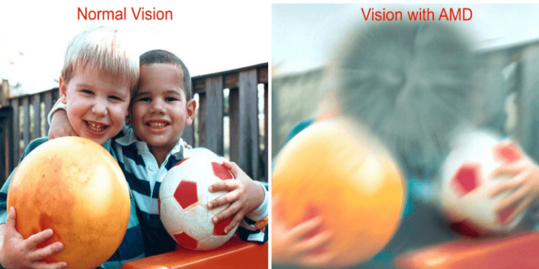
2. AGE RELATED MACULAR DEGENERATION (ARMD)
Age-related macular degeneration (ARMD) is the most common cause of irreversible vision loss in people over age of 60 years. Cells in the macula degenerate (the central, and most sensitive part of the retina at the back of the eye), that is, they become damaged and die. Damage to the macula affects your central vision which is needed for reading, writing, driving, recognizing people’s faces and doing other fine tasks.
TREATMENTS
There is NO TREATMENT for dry AMD, although high dose multivitamin combination has been shown to decrease the risk of visual loss. There are a few treatment options for wet AMD although the best outcomes occur when this disease is detected early.
Laser
Photodynamic therapy (PDT)
Anti-VEGFs (Lucentis, Avastin, Macugen)
Combinations of the above mentioned
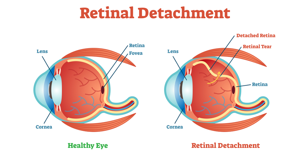
3. RETINAL DETACHMENT
Retinal detachment is separation of the neurosensory retina from the underlying retinal pigment epithelium. The most common cause is a retinal break (a tear or, less commonly, a hole) (rhegmatogenous detachment).
TREATMENTS
When a retinal tear or hole hasn’t yet progressed to detachment, your eye surgeon may suggest one of the following procedures to prevent retinal detachment and preserve vision.
Laser surgery (photocoagulation): The surgeon directs a laser beam into the eye through the pupil. The laser makes burns around the retinal tear, creating scarring that usually “welds” the retina to underlying tissue.
Freezing (cryopexy): After giving you a local anesthetic to numb your eye, the surgeon applies a freezing probe to the outer surface of the eye directly over the tear. The freezing causes a scar that helps secure the retina to the eye wall.
Retinal Surgery as soon as possible to put the retina back in its proper position. The longer the retina stays detached, the less the visual improvement after surgery.
Injecting air or gas into your eye: In this procedure, called pneumatic retinopexy (RET-ih-no-pek-see), the surgeon injects a bubble of air or gas into the center part of the eye (the vitreous cavity). If positioned properly, the bubble pushes the area of the retina containing the hole or holes against the wall of the eye, stopping the flow of fluid into the space behind the retina.
Indenting the surface of your eye: This procedure, called scleral (SKLAIR-ul) buckling, involves the surgeon sewing (suturing) a piece of silicone material to the white of your eye (sclera) over the affected area. This procedure indents the wall of the eye and relieves some of the force caused by the vitreous tugging on the retina.
Draining and replacing the fluid in the eye: In this procedure, called vitrectomy, the surgeon removes the vitreous along with any tissue that is tugging on the retina. Air, gas or silicone oil is then injected into the vitreous space to help flatten the retina. Eventually the air, gas or liquid will be absorbed, and the vitreous space will refill with body fluid. If silicone oil was used, it may be surgically removed months later. Vitrectomy may be combined with a scleral buckling procedure.
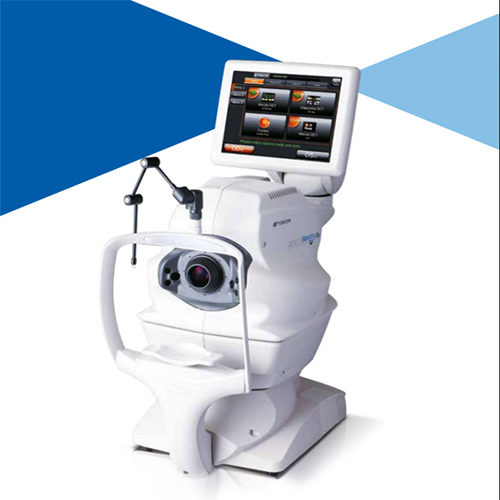
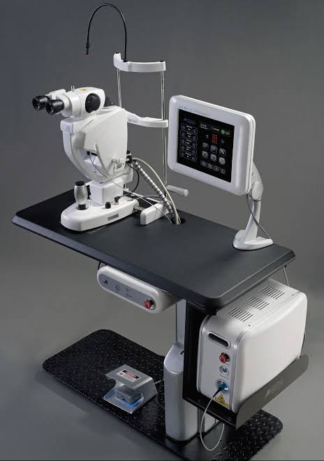
EQUIPMENTS FOR RETINA EVALUATION AND TREATMENT AT OUR HOSPITAL